


Original Article - Year 2014 - Volume 29 -
Diagnosis and treatment for skin cancer in albinos: a descriptive study
Diagnóstico e tratamento do câncer de pele em albinos: estudo descritivo
ABSTRACT
INTRODUCTION: To describe a case series involving albinos as to the form, location, treatment and monitoring of skin tumors.
METHODS: A descriptive, retrospective from April to July 2011, analyzing a total of twelve charts. Seeking the histopathologic results, and descriptions of surgical procedures, collect and report specific data.
RESULTS: Twelve patients had albinos and 273 injuries were studied. Eight men and 4 women, aged between 23 and 80 years, the majority being over 40 years old (92%). The most common injuries were to the head and neck being the most common histological type BCC (36.63%), followed by the CEC. Some injuries were also found tricoblástico carcinoma, high-grade sarcoma, verruca vulgaris, melanoma in situ and Bowen's disease. On average, patients were followed for 98.6 months. Among the surgical procedures the most common was the realization of primary synthesis (82.41%) and second flaps, two microsurgical (VRAM and RALC). The sizes of the lesions was divided into equal or lower than 4 cm (80.20%) and higher than 4 cm (19.80%).
CONCLUSION: Using a short data aggregation was possible to describe a sample with similar data exposed in the international literature, despite the lack of them, allowing a comparison and demonstration about the relationship between albinism and skin tumors, but new series with more patients are needed to better overall evaluation. So prevention remains the best way of monitoring and follow-up of patients with albinism.
Keywords: Albinism; Therapeutics; Carcinoma Basal Cell; Carcinoma Squamous Cel.
RESUMO
INTRODUÇÃO: Descrever a casuística envolvendo albinos, quanto à forma de apresentação, local, tratamento e acompanhamento dos tumores de pele.
MÉTODOS: Estudo descritivo, retrospectivo, de Abril à Julho de 2011, analisando um total de doze prontuários. Buscando nos resultados histopatológicos, e nas descrições dos procedimentos cirúrgicos, reunir e reportar dados específicos.
RESULTADOS: Doze pacientes albinos apresentaram 273 lesões e foram estudados. Oito homens e 4 mulheres, com idades variando entre 23 e 80 anos, sendo a maioria acima de 40 anos (92%). A localização mais comum das lesões foi na cabeça e pescoço, sendo o tipo histológico mais frequente o CBC (Carcinoma Basocelular) (36,63%), seguido do CEC. Algumas lesões também encontradas foram carcinoma tricoblástico, sarcoma de alto grau, verruga vulgar, melanoma in situ e Doença de Bowen. Em média os pacientes foram acompanhados, por 98,6 meses. Dentre os procedimentos cirúrgicos realizados o mais comum foi à realização de síntese primária (82,41%) e em segundo lugar os retalhos, sendo dois microcirúrgicos (VRAM e RALC). Os tamanhos das lesões foram divididos em menores ou iguais a 4 cm (80,20%) e maiores do que 4 cm (19,80%).
CONCLUSÃO: Através de uma breve agregação de dados foi possível descrever uma casuística com dados semelhantes aos expostos na literatura internacional, apesar da escassez dos mesmos, possibilitando uma comparação e demonstração entre a relação sobre albinismo e tumores de pele, porém novas séries descritivas com mais pacientes são necessárias para melhor avaliação global. Assim a prevenção continua sendo a melhor forma de monitoramento e acompanhamento dos pacientes portadores de albinismo.
Palavras-chave: Albinismo; Terapêutica; Carcinoma Basocelular; Carcinoma de Células Escamosas.
Albinism is an autosomal recessive genodermatosis with an incidence that is, in most countries, approximately 1:20,000 individuals, although it is more common in African countries. There are different types of albinism, but the genetic defect is in the synthesis of melanin. The number of melanocytes in the epidermis is normal in most cases, and, in all cases, the formation of melanin is impaired. Some types cause a decrease in tyrosinase, while synthesis activity in others is low. At least 10 different types of oculocutaneous albinism are known1,2. Because of these characteristics, these patients are extremely susceptible to damage caused by sun exposure. They often develop skin lesions, small and benign to large and malignant tumors, with rapid development and high morbidity and mortality rates.
OBJECTIVE
The objective of this study was to describe several cases of albinism. The cases were evaluated for the form of presentation, location, treatment, and monitoring of skin tumors.
METHODS
A retrospective study was conducted over a period of 4 months (April to July 2011) by analyzing the records of 12 patients at the National Cancer Institute. Data was gathered from histopathological results and descriptions of surgical procedures and statistically evaluated.
RESULTS
Twelve albino patients with a total of 273 lesions were investigated. There were 8 men and 4 women (male: female, 2:1) with an age range from 23 to 80 years, although most patients were over 40 years old (92%). (Table 1) The most common lesion location was the head and neck (53.11%), as seen in Figure 1, and the most common histological type was basal cell carcinoma (BCC, 36.63%), as seen in Figure 2, followed by squamous cell carcinoma (SCC, 34.43%). Other lesions found were trichoblastic carcinoma, high-grade sarcoma, verruca vulgaris, melanoma in situ, and Bowen disease. On average, patients have been monitored for 98.6 months so far.
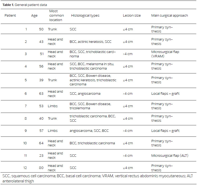
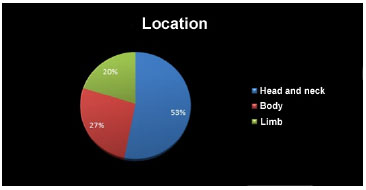
Figure 1. Location of the lesions
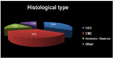
Figure 2. Histological type
Among the surgical procedures performed, the most frequent was primary closure (82.41%), followed by both vertical rectus abdominis myocutaneous (VRAM) and anterolateral thigh (ALT) microsurgical flaps (12%). The size of the lesions were divided into those <4 cm (80.20%) and >4 cm (19.80%). The demographics of the cases studied are summarized in Table 1. One death occurred owing to vascular invasion in the common carotid with BCC, leading to profuse bleeding in a 23-year-old patient. An example of long-term outcome is shown in Figure 3.
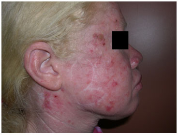
Figure 3. Long-term post-operative outcome of a bilobed flap on the right hemiface
DISCUSSION
According to data in the literature, especially studies from Africa, malignant non-melanoma skin lesions are the most common in albino patients. Spinocellular carcinoma accounts for the vast majority of tumors found3,4,5, but the frequencies of basal cell and spinocellular tumors were similar in our analysis, with slightly more basal cell tumor as they have an indolent progression. We also found a large number of lesions <4cm. This shows an intense screening and close monitoring of patients at the National Cancer Institute (INCA). There were fewer cases undergoing major resections, because of a tumor size >4cm (mainly spinocellular), compared to the literature6-11, as cited by Kingsley et al6. Of the 12% of flaps used to repair defects, 2 cases required flaps; 1, VRAM; and 1, ALT, at a microsurgical distance.
Both the global data and INCA agree that the most common primary site is the head and neck. A negligible number of melanoma cases (1/273) showed a pathophysiology of the disease involving the melanin synthesis. In our case study, compared to international data, the vast majority of treatments for non-melanoma lesions in these patients is primary closure, followed by local flaps.
We recognize that albino patients must drastically reduce their sun exposure by shifting to night professions or staying indoors with light protection and appropriate clothing. Myopia and ocular disorders slow the progress of albinos in school, leading them to withdraw from school and undertake activities in open areas. Therefore, socioeconomic policies involving this population are extremely important.
CONCLUSION
Albinos require intense monitoring and guidance on the use of light protection and on the appearance of new lesions. Basal cell and epidermoid carcinomas are extremely important because of their progressive nature. However, advanced lesions, despite being challenging, can still be treated with both grafts and local or, in specific case, microsurgical flaps. Through a brief aggregation of data, it was possible to describe a case study with data similar to what has been shown in the literature, despite the scarcity of reports, allowing a comparison and demonstration of the relationship between albinism and skin tumors. However, further studies with more patients are needed for a better overall assessment. Prevention thus remains the best method for monitoring of patients with albinism.
REFERÊNCIAS
1. Sampaio,PSA. Dermatologia. In: Sampaio, S, Rivitti, E) editores. Discromias, 3ª ed, São Paulo: Artes Médicas; 2007.Pp.35355.
2. Fitz P, Thomas B. Color Atlas and synopsis of clinical dermatology. In: Fitz P, Thomas B. Color Atlas and synopsis of clinical dermatology. In: Fitz P, T, Wolff, K, Johnson,R (Eds) Pigmentary Disorders, 6th ed, 2009, p.341-344.
3. Ramalingam VS, Sinnakirouchenan R, Thappa DM. Malignant transformation of actinic keratoses to squamous cell carcinoma in an albino. Indian J Dermatol. 2009;54:46-48.
4. Asuquo ME, Ebughe G. Cutaneous cancers in Calabar, Southern Nigeria. Dermatol Online J. 2009;15:11.
5. Diepgen TL, Mahler V. The epidemiology of skin cancer. Br J Dermatol. 2002;146(61):1-6 .
6. Kingsley, O, Bernard, CJ. Skin cancers in albinos in a teaching Hospital in eastern Nigeria - presentation and challenges of care. World J Surg Oncol. 2010;8:73.
7. Berger E. Hunt R. Tzu J. Patel R. Sanchez M. Squamous-cell carcinoma in situ in a patient with oculocutaneous albinism. Dermatol Online J. 2011;17(10):22.
8. Summers CG, Albinism: classification, clinical characteristics, and recent findings. Optom Vis Sci. 2009;86(6):659-62.
9. Kromberg, JG Castle, D., Zwane, E. M. and Jenkins, T. Albinism and skin cancer in Southern Africa. Clinical Genetics. 1989;36:43-52.
10. Yakubu A, Mabogunje OA. Skin cancer in African albinos. Acta Oncol. 1993;32:621-622.
11. Lookingbill,D.P, Lookingbill,G.L, Leppard B. Actinic damage and skin cancer in albinos in northern Tanzania: Findings in 164 patients enrolled in an outreach skin care program. J Am Acad Dermatol. 1995;32(4):653-8.
1 - Specialist in Plastic Surgery at the Plastic Surgery and Microsurgery Service of the National Institute of Cancer (INCA) and Brazilian Society for Plastic Surgery (SBCP)
2 - Masters from UNICAMP, Staff of the Plastic Surgery and Microsurgery Service of INCA
3 - Plastic Surgeon, Head of the Plastic Surgery Department of INCA
Institute: National Cancer Institute of Brazil (Instituto Nacional do Câncer, INCA), RJ, Paulo.
Corresponding author:
Guilherme Graziosi
National Cancer Institute of Brazil (Instituto Nacional do Câncer)
Praça da Cruz Vermelha, 23, Centro
Postal Addressing Code (CEP): 20230-130 - Rio de Janeiro, RJ
Tel.: (21) 32071085
E-mail: guilhermegraziosi@gmail.com
Article received: May 5, 2012
Article accepted: November 28, 2013
 Read in Portuguese
 Read in English
 PDF PT
 Print
Print
 Send this article by email
 How to Cite
 Mendeley
 Pocket