


Case Report - Year 2026 - Volume 41Issue 1
Surgical Correction of Scleral Show in Robinow Syndrome: A Case Report
Correção cirúrgica da exposição escleral em paciente com síndrome de robinow: Um relato de caso
Thaylandiê Basílio Cavalcante Cruz1 ; João Luiz Bezerra de Menezes Saraiva2,*; Kalyne Saraiva Fontenele de Araújo2; Raíra Marques Oliveira2; Isabela Franco Freire2; Vitor de Vasconcellos Muniz1; Salustiano Gomes de Pinho Pessoa1
ABSTRACT
Robinow syndrome is a rare genetic disorder characterized by craniofacial, genital, and limb anomalies. While several corrective procedures for skeletal and facial deformities have been documented, the surgical correction of eyelid malposition in this syndrome has not been previously reported. We present the case of a 12-year-old male with Robinow syndrome and bilateral scleral show due to lower eyelid deficiency. Surgical correction was performed using a muscle-periosteal flap for lower eyelid reinsertion, combined with bilateral canthopexy. Postoperative evaluation demonstrated significant functional and aesthetic improvement, with adequate eyelid apposition to the globe and resolution of scleral exposure. The surgical technique, though established for other eyelid deformities, had not been previously applied in Robinow syndrome. This report documents the first known case of successful surgical correction of scleral show in this condition, demonstrating the feasibility and functional benefits of the surgical approach adopted in this unique clinical context.
Keywords: enetic disorders; reconstructive surgical procedure; plastic surgery; eyelid diseases; case reports; Robinow syndrome
RESUMO
A síndrome de Robinow é uma doença genética rara caracterizada por anomalias craniofaciais, genitais e de membros. Embora diversos procedimentos corretivos para deformidades esqueléticas e faciais tenham sido documentados, a correção cirúrgica da malposição palpebral nessa síndrome não havia relatada anteriormente. Apresentamos o caso de um paciente do sexo masculino, de 12 anos, com síndrome de Robinow e exposição escleral (scleral show) bilateral devido à deficiência da pálpebra inferior. A correção cirúrgica foi realizada por meio de retalho mioperiosteal para reinserção da pálpebra inferior combinado com cantopexia bilateral. A avaliação pósoperatória evidenciou melhora funcional e estética significativa, com adequada aposição palpebral ao globo ocular e resolução da exposição escleral. A técnica cirúrgica, embora consagrada para outras deformidades palpebrais, não havia sido previamente aplicada na síndrome de Robinow. Este relato documenta o primeiro caso descrito de correção cirúrgica bem-sucedida de exposição escleral nesta condição, demonstrando a viabilidade e os benefícios funcionais da abordagem cirúrgica adotada nesse contexto clínico único.
Palavras-chave: doenças genéticas; procedimentos cirúrgicos reconstrutivos; cirurgia plástica; doenças palpebrais; relatos de casos; síndrome de Robinow
Introduction
Robinow syndrome is a rare hereditary disorder characterized by a constellation of features including mesomelic dwarfism, genital hypoplasia, and craniofacial and limb deformities. It presents in two genetic forms: a more severe and prevalent autosomal recessive type, linked to the ROR2 gene, and an autosomal dominant type, associated with the WNT5A gene. Due to significant clinical heterogeneity and the limited number of reported cases, defining its precise phenotypic spectrum remains a challenge.1 The diagnosis is established clinically based on its suggestive features and confirmed through molecular genetic testing that identifies causative mutations.2
The characteristic craniofacial dysmorphism is often pronounced in early childhood and includes macrocephaly, prominent frontal bossing, midface hypoplasia, and significant ocular hypertelorism. This facial structure often results in prominent eyes with an appearance of exophthalmos due to lower eyelid hypoplasia. While surgical management in Robinow syndrome is multidisciplinary, typically addressing deformities such as cleft lip, syndactyly, and genital anomalies, procedures for eyelid malposition have not been previously described in this population.3
We herein report the first documented case of successful surgical correction for scleral show in a patient with Robinow syndrome.
Case Description
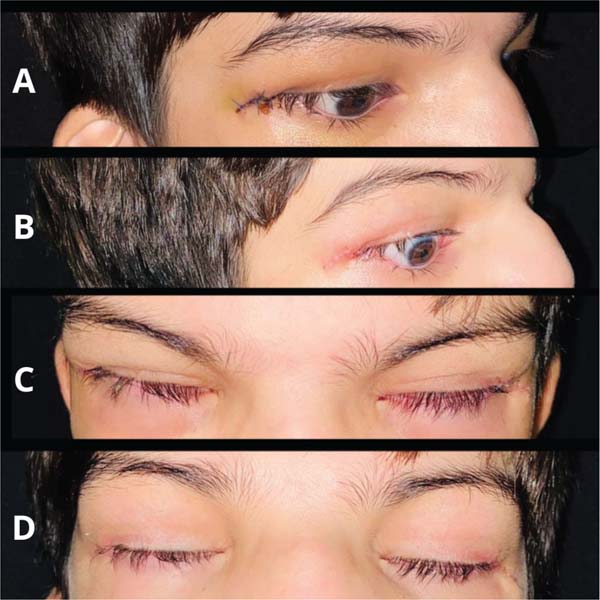
A 12-year-old male with a known diagnosis of Robinow syndrome presented to our department for management of bilateral scleral show. His examination was notable for marked pseudo-exophthalmos secondary to midface hypoplasia and lower eyelid retraction. Given these findings, a surgical plan was formulated to perform lower eyelid reinsertion using a muscle-periosteal flap combined with bilateral canthopexy.
His craniofacial morphology was consistent with the classic phenotype of Robinow syndrome, featuring prominent midface hypoplasia and marked ocular hypertelorism, which contributed to his ocular presentation. To protect patient identity, photographic documentation in this report is limited to the periocular region.
The patient’s past medical history was significant for a complicated neonatal course with anoxia, requiring a three-month admission to a neonatal intensive care unit, and a corneal transplant at age three. A complete history was difficult to obtain due to fragmented medical records. However, his history was suggestive of a congenital heart defect (under pediatric cardiology follow-up) and a possible repaired tracheoesophageal fistula, though the family could not confirm these diagnoses.
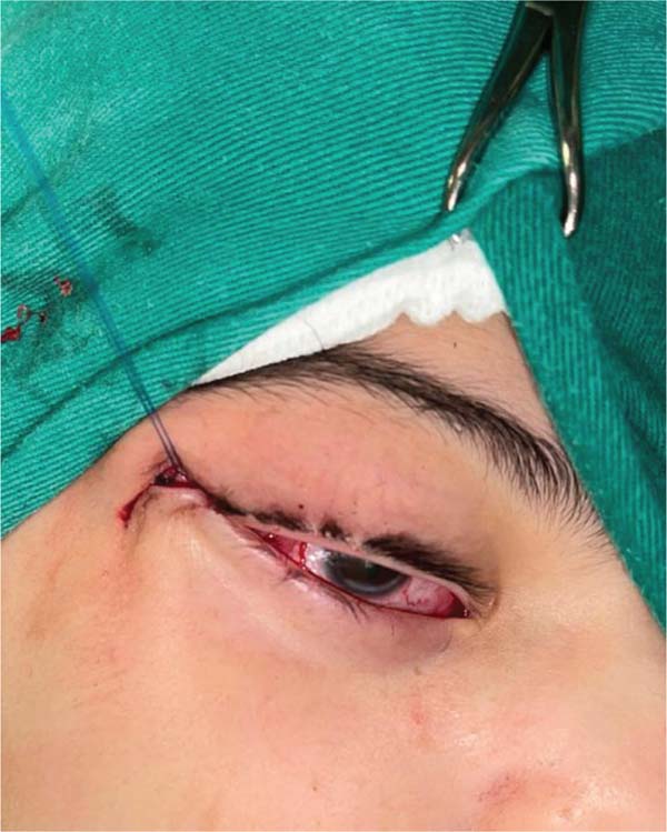
The procedure was performed under local anesthesia with 2% lidocaine. A lateral canthotomy and inferior cantholysis of the lateral canthal tendon were carried out to release the lower eyelid. A tarsal strip was then fashioned from the lateral lower eyelid tarsus and anchored to the periosteum of Whitnall’s tubercle using a 5-0 Prolene suture, providing horizontal eyelid tightening. For vertical support, a myocutaneous flap of the lower eyelid, including the orbicularis oculi muscle, was elevated and secured to the deep temporal fascia (► Fig. 1). The orbicularis muscle layer was then reapproximated using 5-0 Vicryl sutures. Following meticulous hemostasis, the lower eyelid was positioned with its margin at the level of the inferior iris, resulting in 2 mm of superior iris coverage. The canthotomy incision was then closed in anatomical layers (► Fig. 2).
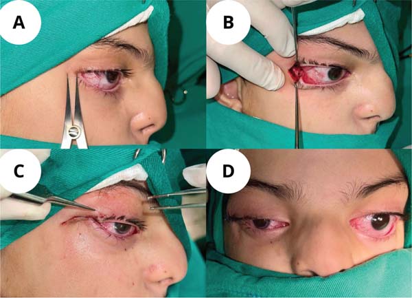
The patient’s postoperative course was uneventful. At the 7-day follow-up visit, the surgical incisions were healing well, with no signs of infection, inflammation, or suture dehiscence. A significant functional improvement was noted, with the patient able to achieve complete eyelid closure and resolution of the preoperative scleral show (► Fig. 3).
The study protocol was reviewed and approved by the Research Ethics Committee of the Hospital Universitário Walter Cantídio -Universidade Federal do Ceará (Approval number: 7.460.061; CAAE registration: 87059225.2.0000.5045).
Discussion
This report details the successful surgical correction of complex lower eyelid malposition in a patient with Robinow syndrome. The patient’s phenotype, including significant midface hypoplasia and ocular hypertelorism, directly contributed to the scleral show,2,4 yet no prior reports have described a surgical correction for this specific deformity.
To our knowledge, this is the first report describing the specific surgical correction of scleral show in a patient with Robinow syndrome. While the surgical technique employed (combining bilateral canthopexy with a muscle-periosteal flap) is an established strategy for other eyelid deformities,5–7 its application in this syndrome had not been previously documented in the literature.
This surgical approach was adopted for its proven ability to correct scleral show, protect the ocular surface, and improve eyelid function. The technique is a well-established strategy for repositioning deficient lower eyelids and preventing subsequent ophthalmologic complications.5,8,9 However, due to the syndrome’s rarity, the management of these features remains challenging, and decisions regarding reconstructive techniques must be tailored to the patient’s anatomical characteristics.
The successful outcome in this case underscores the necessity of a multidisciplinary approach, involving collaboration between plastic surgery, ophthalmology, and genetics, to manage the multi-system manifestations of this syndrome.2,10 The surgical technique was designed to provide durable restoration of eyelid function by anchoring the orbicularis muscle flap to stable periocular structures. The uneventful postoperative course, marked by significant functional improvement, supports the efficacy of this approach in the present patient.
In patients with multiple congenital anomalies, surgical interventions must be prioritized based on functional deficits. In this case, correcting the palpebral malposition was a primary concern due to its implications for corneal protection and preservation of visual function.
This study has several limitations. First, the rarity and the phenotypic variability of Robinow syndrome precludes the generalization of our surgical strategy to all affected individuals. Second, the lack of long-term follow-up data prevents a comprehensive assessment of the correction’sdurability. Finally, the patient’s incomplete medical history limited our ability to fully assess comorbidities that could influence long-term therapeutic planning.
This case report documents the successful application of a planned surgical approach for lower eyelid correction in a patient with Robinow syndrome. The favorable short-term functional outcomes, achieved without complications, support the feasibility and effectiveness of the employed technique in this rare clinical context. Furthermore, this case reinforces the fundamental importance of integrated, multidisciplinary management for patients with complex multi-system syndromes.
REFERENCES
1. Abu-Ghname A, Trost J, Davis MJ, et al. Extremity anomalies associated with Robinow syndrome. Am J Med Genet A 2021; 185(12):3584–3592
2. Roifman M, Brunner H, Lohr J, Mazzeu J, Chitayat D. Autosomal Dominant Robinow Syndrome. In: Adam MP, Feldman J, Mirzaa GM, Pagon RA, Wallace SE, Amemiya A, eds. GeneReviews®. Seattle (WA): University of Washington, Seattle; January 8, 2015
3. Bacino CA. ROR2-Related Robinow Syndrome. In: Adam MP, Feldman J, Mirzaa GM, Pagon RA, Wallace SE, Amemiya A, eds. GeneReviews®. Seattle (WA): University of Washington, Seattle; July 28, 2005
4. Mazzeu JF, Pardono E, Vianna-Morgante AM, et al. Clinical characterization of autosomal dominant and recessive variants of Robinow syndrome. Am J Med Genet A 2007;143(04):320–325
5. Amer AA, Abdellah MM, Hassan NHF, Mounir A. Surgical outcome of epicanthus and telecanthus correction by C-U medial canthoplasty with lateral canthoplasty in treatment of Blepharophimosis syndrome. BMC Ophthalmol 2022;22(01):226
6. Chae SW, Yun BM. Cosmetic Lateral Canthoplasty: Lateral Canthoplasty to Lengthen the Lateral Canthal Angle and Correct the Outer Tail of the Eye. Arch Plast Surg 2016;43(04):321–327
7. Kim YJ, Lee KH, Choi HL, Jeong EC. Cosmetic Lateral Canthoplasty: Preserving the Lateral Canthal Angle. Arch Plast Surg 2016;43 (04):316–320
8. Alencar Filho JIPD, Frota-Júnior JAG, Cardoso GP, Muniz VDV, Alcântara FSD, Pessoa SGDP. Blepharoplasty with canthopexy in a patient with cutis laxa: A case report. Rev Bras Cir Plást 2024;39 (01):e0761
9. Vieira R, Pinho A, Brinca A. Repair of the lower eyelid with primary closure with lateral canthopexy and elevation of the suborbicularis oculi fascia: a simple technique to avoid postoperatory ectropion. Surg Cosmet Dermatol 2018;10(04):1–5
10. Lima AR, Ferreira BM, Zhang C, et al. Phenotypic and mutational spectrum of ROR2-related Robinow syndrome. Hum Mutat 2022; 43(07):900–918
1. Department of Plastic Surgery, Walter Cantídio University Hospital, Federal University
of Ceará (HUWC/UFC), Fortaleza, Ceará, Brazil
2. Health Sciences Center, University of Fortaleza (UNIFOR), Fortaleza, Ceará, Brazil
Study was conducted at Serviço de Cirurgia Plástica e Microcirurgia, Hospital Universitário Walter Cantídio, Fortaleza, Ceará, Brazil.
Compliance with Ethical Standards The study protocol was reviewed and approved by the Research Ethics Committee of the Hospital Universitário Walter Cantídio -Universidade Federal do Ceará (Approval number: 7.460.061; CAAE registration: 87059225.2.0000.5045).
Consent Statement Written informed consent was obtained from a legally authorised representative for the publication of the case report and images in this article.
Financial Support The author(s) received no financial support for the research, authorship, and/or publication of this article.
Data Availability Data will be available upon request to the corresponding author.
Address for correspondence João Luiz Bezerra de Menezes Saraiva, Av. Washington Soares, 1321, CEP 60811-905, Fortaleza, Ceará, Brazil (e-mail: joaoluizbmsaraiva@edu.unifor.br).
Article received: December 17, 2025.
Article accepted: April 10, 2026.
Conflicts of Interest The author(s) declared no potential conflicts of interest with respect to the research, authorship, and/or publication of this article.
 Read in Portuguese
 Read in English
 PDF PT
 Print
Print
 Send this article by email
 How to Cite
 Mendeley
 Pocket
 Facebook
 Twitter