


Original Article - Year 2026 - Volume 41Issue 1
Complications in the Surgical Treatment of Mandibular Condyle Fractures - A Prospective Study
Complicações no tratamento cirúrgico das fraturas de côndilo mandibular - Um estudo prospectivo
José Mauro de Oliveira Squarisi1, ; Lucas Gomes Patrocínio2; Marcell de Mello Naves3; Luna Karla Neves Melo1; Luma de Oliveira Morais1; Marco Túlio Solano Matos1; Maria Mirelle Ferreira Leite Barbosa1
ABSTRACT
Introduction Mandibular fractures are among the most common injuries in the craniofacial region, with involvement of the condylar region reported in 19 to 52% of the cases. Although non-surgical management remains widely used, long-term followup has demonstrated suboptimal control of temporomandibular joint dysfunction and related complications, including chronic pain, ankylosis, condylar arthritis, malocclusion, hypomobility, and facial asymmetry. Although the adoption of the surgical treatment has been increasing in recent years, few contemporary studies have evaluated its complications in condylar fractures.
Materials and Methods The current prospective study was conducted between July 2022 and December 2024, and it included adult patients (≥ 18 years) with mandibular condyle fractures who underwent computed tomography (CT) scans, agreed to the treatment proposed by the clinical team, and were followed up throughout the study period.
Results The study included 33 patients, 23 (69.70%) of whom underwent surgical treatment, and 10 (30.30%), non-surgical management. The overall incidence of complications was of 25.92%, with peripheral facial paralysis and sialocele being the most frequent (11.11% each), followed by temporomandibular joint dysfunction (3.70%).
Conclusion Although the indication for surgical treatment of condylar fractures has been increasing, awareness and careful evaluation of its complications remain essential.
Keywords: mandibular condyle; maxillomandibular fractures; postoperative complications
RESUMO
Introdução Fraturas mandibulares estão entre as lesões mais comuns na região craniofacial, com relato de acometimento da região condilar em 19 a 52% dos casos. Embora o tratamento não cirúrgico ainda seja amplamente utilizado, o acompanhamento no longo prazo tem demonstrado controle subótimo da disfunção da articulação temporomandibular e das complicações relacionadas, incluindo dor crônica, anquilose, artrite condilar, má oclusão, hipomobilidade e assimetria facial. Apesar da crescente adoção do tratamento cirúrgico nos últimos anos, poucos estudos contemporâneos avaliaram suas complicações em fraturas condilares.
Materiais e Métodos Este estudo prospectivo foi conduzido entre julho de 2022 e dezembro de 2024 e incluiu pacientes adultos (≥ 18 anos) com fraturas do côndilo mandibular que foram submetidos à tomografia computadorizada (TC), concordaram com o tratamento proposto pela equipe clínica e foram acompanhados durante todo o período.
Resultados O estudo incluiu 33 pacientes, 23 (69,70%) dos quais foram submetidos ao tratamento cirúrgico, e 10 (30,30%), ao tratamento não cirúrgico. A incidência geral de complicações foi de 25,92%, sendo a paralisia facial periférica e a sialocele as mais frequentes (11,11% cada), seguidas por disfunção da articulação temporomandibular (3,70%).
Conclusão Embora a indicação para o tratamento cirúrgico de fraturas condilares tenha aumentado, a conscientização e a avaliação cuidadosa de suas complicações continuam sendo essenciais.
Palavras-chave: côndilo mandibular; fraturas maxilomandibulares; complicações pós-operatórias
Introduction
Mandibular fractures are among the most common fractures in the craniofacial region. Involvement of the condylar region occurs in 19% to 52% of these fractures, which are associated with functional abnormalities, such as malocclusion, trismus, chronic pain, temporomandibular joint dysfunction (TMD, which reduces mouth opening), and facial asymmetry.1,2
The therapeutic options include non-surgical management (involving maxillomandibular fixation, a restricted liquid diet, early physiotherapy, and orthodontic control with elastics) and surgery, which involves open reduction with internal fixation using miniplates and screws.3,4
Historically, the conservative treatment has been widely adopted because of concerns regarding surgical complications, such as unesthetic scarring, peripheral facial paralysis, infection, salivary fistula, and sialocele. However, the long-term follow-up of conservatively-managed patients has demonstrated suboptimal control of TMD, chronic pain, temporomandibular joint ankylosis, condylar arthritis, malocclusion, hypomobility, and facial asymmetry. Consequently, the surgical treatment has been increasingly adopted in recent years.1,5–9
Although several studies have evaluated the outcomes of the conservative and surgical managements, few have specifically assessed the complication rates associated with the surgical treatment.3,10–14
Objective
The present study aimed to evaluate the epidemiological profile and incidence of complications in patients with condylar fractures undergoing surgical treatment at a tertiary general hospital.
Materials and Methods
The current prospective study was conducted from July 2022 to December 2024, and it included patients with mandibular condyle fractures treated by the cranio-maxillofacial surgery team at the Teaching Hospital of Universidade Federal de Uberlândia.
The inclusion criteria were age ≥ 18 years, performance of computed tomography with three-dimensional reconstruction (3D CT), and the patient’s agreement with the treatment proposed by the medical team (whether surgical or non-surgical). Patients younger than 18 years or those who did not complete a minimum of 6 months of posttrauma outpatient follow-up by the end of the study were excluded. The institutional Ethics in Research Committee approved the study under protocol number CAAE 57095822.0.0000.5152.
Results
We monitored 33 patients with fractures involving the mandibular condyle who met the inclusion criteria. The age of the patients, most of whom were male individuals, ranged from 18 to 87 (mean: 43.7) years (►Table 1 ). The most common trauma mechanisms were motor vehicle accidents, followed by falls and physical assault (►Table 1 ). Condylar fractures were unilateral in 87.88% of the patients, and bilateral in 12.12% (►Table 2 ).
| Data | Number of cases | Percentage |
|---|---|---|
| Sex | ||
| Male | 25 | 75.75% |
| Female | 8 | 24.24% |
| Total | 33 | |
| Trauma mechanism | ||
| Motor vehicle accident | 14 | 42.40% |
| Fall | 10 | 30.30% |
| Physical assault | 9 | 27.26% |
| Total | 33 |

| Fracture type | Number of cases | Percentage |
|---|---|---|
| Unilateral | 29 | 87.88% |
| Bilateral | 4 | 12.12% |
| Total | 33 |

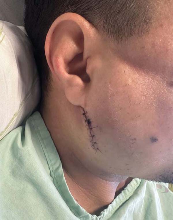
Surgical treatment was indicated for patients with fragment deviations greater than 45° and loss of mandibular ramus height exceeding 2 mm, as confirmed by thin-slice 3D CT. This approach was selected for 69.70% of the patients (►Table 3 ). The most frequently used approach was the transparotid retromandibular approach, followed by the preauricular approach for high condylar fractures (►Table 4 ; ►Figs. 1 –2 ).
| Treatment type | Number of cases | Percentage |
|---|---|---|
| Surgical | 23 | 69.70% |
| Non-surgical | 10 | 30.30% |
| Total | 33 |

| Approach routes | Number of cases | Percentage |
|---|---|---|
| Retromandibular | 18 | 66.67% |
| Preauricular | 9 | 33.34% |
| Total | 27 |

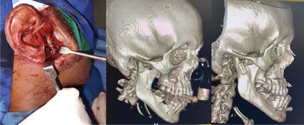
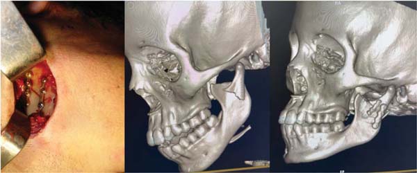
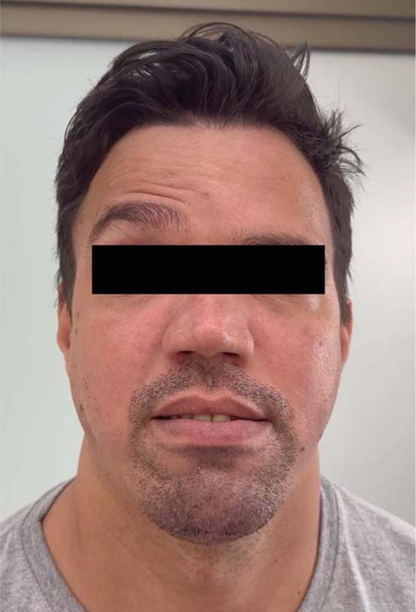
We observed 7 complications: 3 cases of peripheral facial paralysis (2 which resolved completely within 3 months, and 1 that resulted in permanent paralysis of the unilateral frontal branch), three cases of sialocele (1 which improved with drainage and compressive dressing, and 2 that improved after a single session of botulinum toxin), and 1 case of TMD with reduced oral opening (►Figs. 3 –4 ). The overall incidence of complications was of 25.92%, with sialocele and peripheral facial paralysis each occurring in 11.11% of the cases. The incidence of TMD was of 3.70% (►Table 5 ).
| Complications | Number of cases | Percentage |
|---|---|---|
| Peripheral facial paralysis | 3 | 11.11% |
| Sialocele | 3 | 11.11% |
| Temporomandibular joint dysfunction | 1 | 3.70% |
| Total | 7 | 25.92% |

Discussion
The current study evaluated the incidence of complications in patients with condylar fractures who underwent surgical treatment. Consistent with our study, other authors2,10,12,15 have shown a predominance of condylar fractures in male patients of working age.
Motor vehicle accidents, followed by falls and physical assault, were reported as the main mechanisms of trauma in 3 recent studies2,16,17 that corroborate our findings, though the 2023 study by Tatsumi et al.17 identified falls as the most frequent mechanism. The high prevalence of unilateral fractures observed in our hospital, with a rate of 87.88%, is consistent with previous studies,2,10,13,15 which have reported rates of up to 89.7%.
Most of our patients (69.70%) underwent open reduction and internal fixation. Although a 2023 systematic review by Jazayeri et al.1 reported benefits of the surgical treatment, particularly in patients with significantly-displaced fractures, recent studies2,13,18 indicate that the non-surgical management remains the predominant approach, contrasting with our findings. This discrepancy may be explained by the fact that our hospital is a tertiary care center, to which only the most severe trauma cases are referred.
Consistent with our findings, other studies12,13,15,16 have also reported a predominance of the retromandibular approach over the preauricular approach. More recently, some centers have begun to adopt the intraoral approach as an alternative to cutaneous techniques.
Peripheral facial paralysis may occur in transient or permanent forms. Its mechanism can involve compression from surgical retractors or direct nerve injury, and it can occur regardless of the surgical approach used. The literature reports an incidence ranging from 2.43% to 18.3%, consistent with our observed rate of 11.11%. Management strategies vary according to the cause and chronicity of the paralysis, typically involving multidisciplinary follow-up. Treatments range from less invasive options, such as botulinum toxin, to complex microsurgical procedures, which may include regional muscle transfers and cross-face nerve grafts.10,12,13,18,19
Sialocele is associated with the retromandibular surgical route, in which the fracture is approached through the parotid gland. It occurred in 11.11% of our patients, compared with the rate of 15.8% reported by Kulkarni et al.12 Management options for this complication include repeated aspirations, external compression, antisialagogue medications, botulinum toxin, radiation therapy, and, in rare cases, parotidectomy.12
Several studies, including the meta-analysis by Jazayeri et al.,1 have evaluated TMD following condylar fractures, comparing non-surgical management with open reduction and internal fixation. The surgical treatment often yielded superior outcomes in terms of temporomandibular joint pain, lateral mandibular movements, and malocclusion. To the best of our knowledge, no research has compared different surgical approaches and their impact on TMD incidence. In the current study, TMD with reduced postoperative oral opening only occurred in 3.70% of the patients, whereas other authors1,13,18 have reported rates ranging from 10.8% to 17.1%; however, our sample size was smaller than those of previous reports.
The limitations of the present study include the relatively small sample size compared with larger specialized centers on cranio-maxillofacial trauma, as well as the potential for referral bias, since less complex cases may not be directed to our service, resulting in a higher proportion of severe presentations.
Conclusion
Although the indication for surgical treatment of mandibular condyle fractures is increasing, it carries the risk of postoperative complications that require recognition for their prevention or to establish the appropriate management.
REFERENCES
1. Jazayeri HE, Lopez J, Khavanin N, et al. Comparative Benefits of Open versus Closed Reduction of Condylar Fractures: A Systematic Review and Meta-Analysis. Plast Reconstr Surg 2023;151(04): 664e–672e. Doi: 10.1097/PRS.0000000000010009
2. Franke A, Matschke JB, Weiland B, et al. A single-centre retrospective 10-year experience of the rhombic 3D condylar fracture plate for open reduction and internal fixation of condylar neck and base fractures. J Craniomaxillofac Surg 2024;52(05): 622–629. Doi: 10.1016/j.jcms.2024.03.001
3. Gilliland J, Ritto F, Tiwana P. Complications of the Transmasseteric Anteroparotid Approach for Subcondylar Fractures: A Retrospective Study. Craniomaxillofac Trauma Reconstr 2022;15(01): 66–71. Doi: 10.1177/19433875211016923
4. Fuentes CC, Mori FA, Pérez SV, et al. Clinical Experience With a Less Invasive Surgical Transparotid Approach and Trapezoidal Plate for Neck and Base Condylar Fractures: A Retrospective Study. Craniomaxillofac Trauma Reconstr 2024;17(04):NP44–NP50. Doi: 10.1177/19433875241242938
5. De Riu G, Gamba U, Anghinoni M, Sesenna E. A comparison of open and closed treatment of condylar fractures: a change in philosophy. Int J Oral Maxillofac Surg 2001;30(05):384–389. Doi: 10.1054/ijom.2001.0103
6. Ellis E III, Throckmorton GS. Treatment of mandibular condylar process fractures: biological considerations. J Oral Maxillofac Surg 2005;63(01):115–134. Doi: 10.1016/j.joms.2004.02.019
7. Landes CA, Lipphardt R. Prospective evaluation of a pragmatic treatment rationale: open reduction and internal fixation of displaced and dislocated condyle and condylar head fractures and closed reduction of non-displaced, non-dislocated fractures. Part I: condyle and subcondylar fractures. Int J Oral Maxillofac Implants 2005;34(08):859–870. Doi: 10.1016/j.ijom.2005.04.021
8. Nussbaum ML, Laskin DM, Best AM. Closed versus open reduction of mandibular condylar fractures in adults: a meta-analysis. J Oral Maxillofac Surg 2008;66(06):1087–1092. Doi: 10.1016/j. joms.2008.01.025
9. Liu Y, Bai N, Song G, et al. Open versus closed treatment of unilateral moderately displaced mandibular condylar fractures: a meta-analysis of randomized controlled trials. Oral Surg Oral Med Oral Pathol Oral Radiol 2013;116(02):169–173. Doi: 10.1016/j.oooo.2013.02.023
10. Girhe V, Patil V, Bhujbal R, Singh R, Dewang P, Vaprani G. Pre- auricular Transparotid Approach for the Management of Mandibular Condylar Fracture: An Experience of 82 Cases. J Maxillofac Oral Surg 2022;21(03):916–922. Doi: 10.1007/s12663-021-01565-6
11. Lu XW, Liu XG. Risk of complications with retromandibular transparotid vs. anteroparotid approach for condylar fractures: a systematic review and meta-analysis. Eur Rev Med Pharmacol Sci 2022;26(03):799–809. Doi: 10.26355/eurrev_202202_27988
12. Kulkarni V, Chowdhury SKR, Ghosh S, Rajkumar K. Incidence of Facial Nerve Injury and Sialocele Formation Following Mandibular Condylar and Sub-condylar Fracture Fixation. J Maxillofac Oral Surg 2024;23(02):380–386. Doi: 10.1007/s12663-021-01674-2
13. Kumar M, Ramanujam S, Kumaravelu R, Cheeman RS, Periera RJ, Titus S. Open vs Closed Management of Condylar Fracture Our Experience of 100 Cases in a Suburban Tertiary Care Hospital. Craniomaxillofac Trauma Reconstr 2024;17(01):4–12. Doi: 10.1177/19433875221143852
14. Bicsák Á, Koch L, Claussen J, et al. Long-term evaluation of complications after osteosynthesis of the jaws in patients with head and neck trauma: an analysis from a German highest level trauma center 2007-2023. Sci Rep 2025;15(01):11333. Doi: 10.1038/s41598-025-95455-3
15. Mansuri Z, Dhuvad J, Anchlia S, Bhatt U, Rajpoot D, Patel H. Comparison of three different approaches in treatment of mandibular condylar fractures - Our experience. Natl J Maxillofac Surg 2023;14(02):256–263. Doi: 10.4103/njms.njms_485_21
16. Sinha A, Natarajan S. Comparative Evaluation of Clinical and Radiological Outcomes of Retromandibular Transparotid and Transoral Endoscopic-Assisted Approach for Surgical Management of Mandibular Subcondylar Fractures. Cranio-maxillofac Trauma Reconstr 2021;14(02):90–99. Doi: 10.1177/1943387520949099
17. Tatsumi H, Matsuda Y, Toda E, Okui T, Okuma S, Kanno T. Postoperative Complications following Open Reduction and Rigid Internal Fixation of Mandibular Condylar Fracture Using the High Perimandibular Approach. Healthcare (Basel) 2023;11(09):1294. Doi: 10.3390/healthcare11091294
18. Nys M, Van Cleemput T, Dormaar JT, Politis C. Long-term Complications of Isolated and Combined Condylar Fractures: A Retrospective Study. Craniomaxillofac Trauma Reconstr 2022;15(03): 246–252. Doi: 10.1177/19433875211026759
19. Viterbo F, Menezes BFd. Harvest of fascia lata for use in a temporal orthodromic flap in the treatment of facial paralysis. Rev Bras Cir Plást 2022;37(04):518–522. Doi: 10.5935/2177-1235.2022RBCP.620-en
1. Department of Cranio-Maxillofacial Surgery, Hospital das Clínicas, Universidade
Federal de Uberlândia (EBSERH), Uberlândia, MG, Brazil
2. Clínica Otoface, Uberlândia, MG, Brazil
3. Clínica Rhinocenter, Uberlândia, MG, Brazil
Data Availability Data will be available upon request to the corresponding author.
Financial Support The authors declare that they did not receive financial support from agencies in the public, private or nonprofit sectors to conduct the present study.
Address for correspondence José Mauro de Oliveira Squarisi, MD, MSc, PhD, Departamento de Cirurgia Craniomaxilofacial, Hospital das Clínicas, Universidade Federal de Uberlândia (EBSERH), Uberlândia, MG, Brazil (e-mail: josemauro@ufu.br ).
Article received: June 26, 2025.
Article accepted: November 24, 2025.
Conflict of interests The authors have no conflict of interests to declare.
Editor-in-Chief: Dov Charles Goldenberg.
 Read in Portuguese
 Read in English
 PDF PT
 Print
Print
 Send this article by email
 How to Cite
 Mendeley
 Pocket
 Facebook
 Twitter