


Case Report - Year 2026 - Volume 41Issue 1
Early Reconstruction of Severe Aplasia Cutis Congenita Using Opposing Flaps: First Case Reported in Colombia
Reconstrução precoce de aplasia cutânea congênita grave utilizando retalhos opostos: Primeiro caso relatado na Colômbia
Darlyn Dayana Castillo Diaz1, ; Pedro Fabián López Aldana2; Juan Dario Alviar Rueda2
ABSTRACT
Aplasia cutis congenita (ACC) is a rare congenital defect characterized by localized absence of skin, most commonly on the scalp. Severe cases may involve bone and meningeal exposure, posing a high risk of infection, bleeding, and neurological complications. We herein present a case of a full-term newborn diagnosed with severe ACC over the sagittal suture, involving the skin, the subcutaneous tissue, and the parietal bone with an exposed and intact dura mater. The patient underwent early surgical reconstruction on day 3 of life using semicircular opposing flaps to achieve complete defect coverage. Follow-up at 24 days revealed a well-healed, linear scar with early hypertrophic changes and partial hair regrowth, and no neurological sequelae. This case highlights the importance of early diagnosis, timely surgical intervention with local opposing flaps as a safe and reproducible option, and the essential role of a multidisciplinary team in managing severe ACC, particularly in complex neonatal settings. It represents, to our knowledge, the first reported case in Colombia of a cranial ACC defect with bone and dural exposure successfully treated with local advancement flaps.
Keywords: aplasia cutis congenita; scalp; surgical flaps; congenital abnormalities
RESUMO
A aplasia cutânea congênita (ACC) é uma malformação congênita rara caracterizada pela ausência localizada de pele, emespecial no couro cabeludo. Emcasos graves, pode haver exposição óssea e meníngea, o que representa alto risco de infecção, sangramento e complicações neurológicas. Apresentamos o caso de um neonato a termo diagnosticado com ACC grave sobre a sutura sagital, com acometimento de pele, tecido subcutâneo e osso parietal, além de exposição da dura-máter intacta. O paciente foi submetido à reconstrução cirúrgica precoce no terceiro dia de vida, por meio de retalhos semicirculares opostos para a obtenção de cobertura completa do defeito. O acompanhamento aos 24 dias do procedimento revelou uma cicatriz linear bem regenerada, com alterações hipertróficas iniciais e recrescimento parcial do cabelo, semsequelas neurológicas. Este caso destaca a importância do diagnóstico precoce, da intervenção cirúrgica oportuna com retalhos opostos locais como uma opção segura e reprodutível, e o papel essencial de uma equipe multidisciplinar no manejo da ACC grave, particularmente em contextos neonatais complexos. Representa, pelo que sabemos, o primeiro caso relatado na Colômbia de uma ACC craniana comexposição óssea e dural tratada com sucesso mediante retalhos locais de avanço.
Palavras-chave: aplasia cutânea congênita; couro cabeludo; retalhos cirúrgicos; anomalias congênitas; neonato
Introduction
Aplasia cutis congenita (ACC) is a rare disease characterized by the localized absence of some or all layers of skin at birth. It usually occurs on the scalp,1 although it can also affect other regions such as the face, trunk, or extremities. The worldwide prevalence of ACC is of approximately 1 in every 10 thousand live births,2 and between 20 and 30% of the cases present underlying bone involvement.3 Regarding the specific prevalence in Colombia, there is no concrete data available in the literature. Its etiology is multifactorial and not fully understood, but it includes various genetic factors, intrauterine ischemia, infections, and the use of teratogenic agents and drugs.2 This pathology may occur in isolation or in association with other congenital anomalies. Autosomal dominant inheritance is the most common, but recessive inheritance has also been described.4 The diagnosis is clinical and established during the neonatal physical examination. The Frieden5 classification subdivides ACC into 9 groups based on cause, location, and associated malformations. Treatment depends on various factors, such as size, location, and the degree of involvement of adjacent structures.
On the other hand, newborns with severe aplasia cutis congenita who have exposed meninges or brain tissue are at significantly increased risk of developing serious complications such as neonatal meningitis and sepsis due to the loss of the skin-bone barrier that normally protects the central nervous system (CNS). These infectious conditions, along with the possibility of bleeding or dehydration through the defect, contribute to a high mortality rate, ranging from 20 to 50%, especially in the absence of timely surgical and medical management.6 The objective of the current article is to describe the first case reported in Colombia of a severe ACC treated with scalp opposition flaps, a timely and reproducible surgical treatment strategy.
Case Report
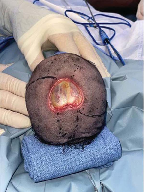
A full-term neonate at 38 weeks was born to a 14-year-old primiparous mother with no relevant medical history. Prenatal ultrasound showed large bowel dilation (14.9 mm), so delivery was scheduled at 38 weeks. Due to poor labor progress, a cesarean section was performed on February 16. The patient was born weighing 2,710 g, measuring 50 cm, with a head circumference of 35 cm, and an appearance, pulse, grimace, activity, and respiration (Apgar) score of 8/9, with no respiratory distress. A physical examination revealed a 3 × 4-cm defect above the sagittal suture, with absent parietal bone, an exposed but intact dura mater, and no herniation or abnormal neurological signs (►Fig. 1). The patient was hemodynamically stable, with no ventilatory or vasopressor requirements. Sterile packing was indicated, along with transfer to the neonatal intermediate care unit (NICU), fasting with 10% dextrose. A multidisciplinary evaluation by plastic surgery, neurosurgery, and genetics was requested. The initial laboratory tests and thoracoabdominal radiograph were unremarkable.
On day 2 of life, plastic surgery recommended surgical closure using local flaps, while neurosurgery advised clinical monitoring, brain magnetic resonance imaging (MRI), and surveillance for intracranial hypertension or cerebrospinal fluid leakage. On day 3, a genetic evaluation described a non-consanguineous couple (14-year-old mother and 18-year-old father). The clinical findings included ACC, a single palmar crease, and fifth-digit clinodactyly. A diagnostic protocol was initiated, including G-banded karyotyping and evaluation of six organ systems (through MRI, echocardiogram, renal ultrasound, spine radiograph, and ophthalmologic and auditory assessments).
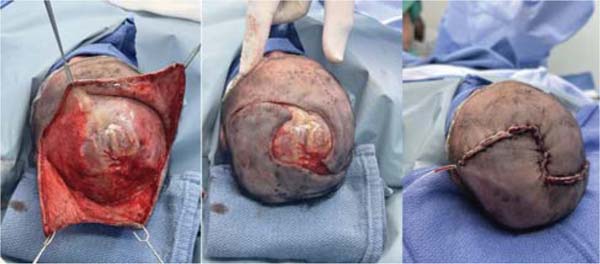
That same day, surgical reconstruction was performed. The intraoperative findings confirmed a severe form of ACC, with absence of skin, subcutaneous tissue, and calvarial bone, along with meningeal exposure. Two semicircular advancement flaps were designed and elevated to achieve complete coverage of the defect (►Fig. 2). Closure was achieved using 4-0 polydioxanone and 4-0 nylon sutures. Postoperative care included cefotaxime (137 mg intravenously [IV] every 12 hours) and ampicillin (274 mg IV every 8 hours), as well as continuous morphine (30 mcg/kg/hour, titrated to 15 mcg/kg/hour) infusion for analgesia.

On postoperative day 1, the infant experienced an isolated episode of abnormal eye movements, spasticity, and cyanosis. Anticonvulsant treatment was initiated with midazolam (0.2 mg/kg/h, 0.5 mg IV) and a loading dose of phenobarbital (20 mg/kg, 54 mg, and then, 7 mg IV every 12 hours). On day two, the Pediatric Ophthalmology Division diagnosed bilateral exposure keratitis and initiated treatment with polyacrylic acid lubricant drops, 1 drop in both eyes 3 times a day for 10 days. Due to clinical deterioration, including fever and a seizure, the antibiotic regimen was escalated to cefotaxime (137 mg IV every 8 hours, 50 mg/kg/dose) and vancomycin (41 mg IV every 12 hours, 15 mg/kg/dose) for 7 days. A lumbar puncture was performed to rule out CNS infection. The cerebrospinal fluid analysis showed normal parameters and negative results on a multiplex array panel for meningitis/encephalitis. Sedation and analgesia were adjusted, and paracetamol replaced morphine as pain control.
Subsequent wound assessments and dressings on days 5, 8, and 12 showed no signs of superinfection or dehiscence. The patient was discharged from plastic surgery for outpatient follow-up every 3 days. By day 11, the ophthalmologic symptoms had resolved, and treatment was discontinued. The patient was weaned off oxygen and transitioned to oral feeding following evaluation by speech therapy.
The newborn was discharged on March 2, 2025, in stable condition, tolerating feeds, with no neurological sequelae. Follow-up appointments were scheduled with general medicine, neonatology, pediatrics, pediatric neurology, neurosurgery, and genetics. A spine radiograph and chromosomal study were pending.
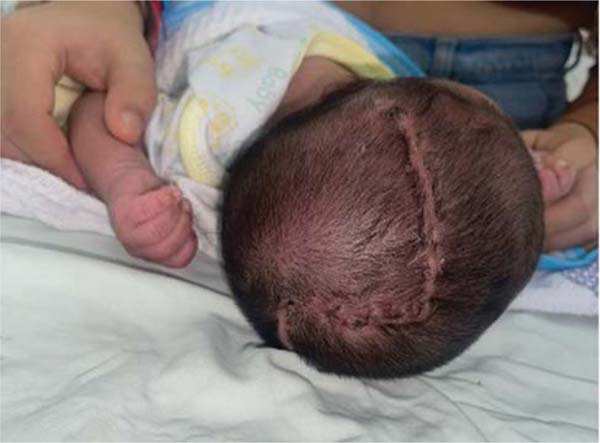
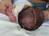
At 24 days postoperatively, the patient exhibited a linear Z-shaped scar in the parieto-occipital region, with early hypertrophic changes and partial hair regrowth, without signs of infection, wound dehiscence, or alopecia (►Fig. 3).
Discussion
We herein present a case of severe ACC with exposed meninges that was successfully closed with opposing flaps. This approach offers a simple and safe technique that can be applied for early reconstruction in severe neonatal presentations of ACC, such as the one herein reported.
A very rare congenital defect manifested by local absence of skin and primarily affects the scalp., the clinical presentation of ACC can vary widely, but the most severe cases are characterized by exposure of deeper structures such as bones and meninges, as evidenced in the case herein reported. This variety in presentation has led to different classifications, but one of the most accepted is the one proposed by Frieden,5 which divides ACC into nine types based on the affected site, the number of lesions, and the association with other congenital anomalies or syndromes (►Table 1).
| Category | Affected body area | Inheritance type |
|---|---|---|
| Group 1: ACC of the scalp without multiple anomalies | Scalp, usually at the vertex | Autosomal dominant or sporadic |
| Group 2: ACC of the scalp with limb anomalies | Midline of the scalp | Autosomal dominant |
| Group 3: ACC of the scalp with epidermal nevi or organoid nevi | Scalp, may be asymmetric | Sporadic |
| Group 4: ACC over embryological malformations | Abdomen, lumbar skin, scalp; any location | Depends on underlying condition |
| Group 5: ACC with fetus papyraceus or associated placental infarcts | Multiple, symmetrical areas, often stellate or linear, on the scalp, thorax, flanks, axillae, and limbs | Sporadic |
| Group 6: ACC associated with epidermolysis bullosa: usually localized blisters, without multiple congenital anomalies | Extremities | Depends on the type of epidermolysis bullosa: can be autosomal dominant or recessive |
| Group 7: ACC localized on limbs, without blisters | Pretibial areas; backs of hands and feet; extensor surfaces of wrists | Autosomal dominant or recessive |
| Group 9: ACC associated with malformation syndromes | Scalp; any location | Variable, depending on the specific syndrome |

In the patient in question, the midline location of the scalp, with bone loss and meningeal exposure, in addition to the finding of two minor anomalies in the extremities, suggests type 2, which may be related to dysfunctional cranial midline formation. No lesions were detected elsewhere in the patient; at the time of the writing of the current article, no additional disease or abnormality had been detected, and other systemic examinations were normal. Furthermore, no drug use during pregnancy was recorded, nor was there any history of trauma. No genetic diseases were detected in the family, nor was there consanguinity between the parents.
Most cases of ACC with small defects without exposure of deeper structures can be managed conservatively, as spontaneous epithelialization is allowed within weeks or months.7 However, in cases such as thie one herein reported, when the defect is extensive or there is bone involvement with meningeal exposure, early surgical management becomes the safest option, because there is a high risk of complications such as bleeding, serious infections (such as meningitis), dehydration, direct trauma to the CNS, and neurological damage.8,9 In this context, early closure with local flaps represents an effective strategy, providing secure coverage, reducing hospitalization time and the risk of infection.
Two cases of ACC with meningeal exposure have been previously reported in Colombia; however, both differ significantly from the case herein presented. The first one10 involved a newborn with type-IV ACC in the dorsolumbar region, associated with hydrocephalus and extensive loss of skin, bone, and dura mater, which required ventriculoperitoneal shunting. The second case11 was that of a 6 × 4-cm cranial defect with bone and meningeal exposure, which was managed conservatively using hydrocolloid dressings, avoiding surgical intervention. In contrast, the case herein presented is, to our knowledge, the first reported in Colombia involving a cranial ACC defect with bone and dural exposure treated successfully with local advancement flaps.
This approach offers a local-coverage strategy with low morbidity, preserving the surrounding tissue and minimizing surgical complexity. Importantly, it circumvents the need for microvascular free flaps, which are technically demanding, resource-intensive, and often not ideal in the neonatal population due to their physiological fragility and limited donor-site availability. Additionally, the technique enabled effective closure of the scalp and calvarial defect while avoiding the need for prosthetic materials or tissue expanders, which are associated with higher risks and complications in neonates.12 Despite the severity of the defect, the patient progressed favorably, without major postoperative complications, supporting this technique as a viable and reproducible alternative in similar cases and in centers with specialized equipment. The case also highlights the value of a multidisciplinary approach. The coordinated involvement of neonatology, plastic surgery, neurosurgery, genetics, infectious disease, ophthalmology, and pediatric neurology was essential for comprehensive diagnosis, timely treatment, and prevention of sequelae. Long-term follow-up is essential to detect late complications, neurodevelopmental disorders, or manifestations of associated syndromes.
A key limitation of the current study is the fact that it is a single case report, which limits the generalizability of our findings. While successful in this instance, further research with a larger patient cohort would be necessary to validate the widespread applicability and efficacy of this surgical technique.
Conclusion
In conclusion, we have reported a rare case of severe ACC with meningeal exposure, with a very favorable clinical evolution due to an early diagnosis and a timely surgical approach. It also highlights the value of the opposing flap technique as an effective, safe, reproducible, and efficient option in complex pediatric settings. However, management must be individualized based on the extent of the lesion and the newborn’s condition, and we reaffirm the importance of early intervention and multidisciplinary planning.
Furthermore, appropriate follow-up by all specialties is essential to promptly detect any complications, continue developmental monitoring, and complete pending examinations. In addition, plastic surgery follow-up is essential to monitor flap development, continue healing, and prevent cosmetic or functional complications.
REFERENCES
1. Evers ME, Steijlen PM, Hamel BC. Aplasia cutis congenita and associated disorders: an update. Clin Genet 1995;47(06): 295–301. Doi: 10.1111/j.1399-0004.1995.tb03968.x
2. Orphanet. Aplasia cutis congenita [Internet]. Paris: INSERM; c1997–2025 [citado 2025 abr 29]. Disponível em: https://www.orpha.net/en/disease/detail/1114?name=aplasia%20cutis%20congenita&mode=name
3. Elliott AM, Teebi AS. Further examples of autosomal dominant transmission of nonsyndromic aplasia cutis congenita. Am J Med Genet 1997;73(04):495–496
4. Online Mendelian Inheritance in Man (OMIM®). APLASIA CUTIS CONGENITA, NONSYNDROMIC; ACC [Internet]. Baltimore, MD: McKusick-Nathans Institute of Genetic Medicine, Johns Hopkins University; c1966–2025 [citado 2025 abr 29]. Disponível em: https://www.omim.org/entry/107600
5. Frieden IJ. Aplasia cutis congenita: a clinical review and proposal for classification. J Am Acad Dermatol 1986;14(04):646–660. Doi: 10.1016/s0190-9622(86)70082-0
6. Bharti G, Groves L, David LR, Sanger C, Argenta LC. Aplasia cutis congenita: clinical management of a rare congenital anomaly. J Craniofac Surg 2011;22(01):159–165. Doi: 10.1097/SCS.0b013e3181f73937
7. Iljin A, Aliev RM. Aplasia cutis congenita of the scalp: Conservative vs. surgical treatment. Ann Plast Surg 2020;84(04):466–470
8. Smith DW, Miller JR. Absence of skin at birth (congenital absence of skin). Am J Dis Child 1963;105:574–579
9. Chitnis A, Nanjappa B. Aplasia cutis congenita: A rare case report and review of literature. Indian J Plast Surg 2015;48(01):90–92
10. Trujillo-Gómez JR, Becerra-Bonilla D, Sierra-González J. Aplasia cutis congénita tipo IV con exposición de meninges. Rev Mex Neuroci 2020;21(01):45–49
11. Montero-Gómez RA, Salazar-Durán SA, Ruiz-Silva JH. Aplasia cutis congénita del cuero cabelludo: reporte de caso y revisión de la literatura. Rev Iber Lat Am Cir Plást 2021;47(02):188–194
12. Pandey A, Dhingra R, Soni N, et al. Surgical reconstruction of aplasia cutis congenita of scalp: Case series and review. J Cutan Aesthet Surg 2019;12(01):38–42
1. Universidad Industrial de Santander, Bucaramanga, Departamento de Santander, Colombia
2. Reconstructive and Esthetic Plastic Surgery Specialization Program, Department
of Surgery, Universidad Industrial de Santander, Bucaramanga, Departamento de Santander,
Colombia
Data Availability Data will be available upon request to the corresponding author.
Financial Support The authors declare that they did not receive financial support from agencies in the public, private, or non-profit sectors to conduct the present study.
Address for correspondence Darlyn Dayana Castillo Diaz, medical student, Universidad Industrial de Santander, Bucaramanga, Departamento de Santander, Colombia (e-mail: darlyncastillomd@gmail.com).
Article received: May 21, 2025.
Article accepted: January 13, 2026.
Conflict of Interests The authors have no conflict of interests to declare.
Editor-in-Chief: Dov Charles Goldenberg.
 Read in Portuguese
 Read in English
 PDF PT
 Print
Print
 Send this article by email
 How to Cite
 Mendeley
 Pocket
 Facebook
 Twitter