


Calendar of Events - Year 2026 - Volume 41Issue 1
Introducing Vertical Torsoplasty
Apresentando a torsoplastia vertical
Luiz Henrique Benetti Favali1 ; Flavio Henrique Mendes2,; Luis Fernando Spagnuolo Brunello2; Mauricio Orel1
ABSTRACT
Introduction Significant weight loss can lead to body contour deformities due to skin and superficial fascial system laxity. Vertical torsoplasty is an innovative technique aimed at correcting dorsal loosens without horizontal or oblique scars, using median vertical tissue resection.
Objective To present the authors' experience in correcting dorsal flaccidity using vertical torsoplasty.
Methods This retrospective study analyzed medical records of patients who underwent vertical torsoplasty between March 2022 and March 2024. The study included patients of all ages and genders with complaints of dorsal flaccidity. The technique involves vertical resection and tissue suspension, with scars camouflaged by the posterior median sulcus.
Results The analysis included 17 patients (16 women, 1 man), with a mean age of 37 years. Most presented flaccidity classified as Pittsburgh 1 or 2. The average surgery time was 90minutes, with no major complications. Minor complications included scar dehiscence and seroma, which were treated conservatively. Patient satisfaction was high, with scars camouflaged in the posterior median sulcus, preserving its good anatomical definition. Discussion Vertical torsoplasty is an effective alternative for treating dorsal flaccidity, with concealed scars and satisfactory aesthetic
Results. Although its tactical philosophy is comparable to that of anchor abdominoplasty, it focuses on the dorsal region and employs inverse tightening and suspension vectors.
Conclusion Vertical torsoplasty is a safe and effective procedure for correcting mild to moderate dorsal laxity. It promotes improved body contour while preserving the anatomical definition of the region.
Keywords: body contouring; plastic surgery procedures; torso; weight loss; lipectomy
RESUMO
Introdução A perda ponderal significativa pode causar deformidades no contorno corporal devido à frouxidão da pele e do sistema fascial superficial. A torsoplastia vertical é uma técnica inovadora que visa corrigir a flacidez dorsal sem cicatrizes horizontais ou oblíquas, utilizando ressecção tecidual vertical mediana.
Objetivo Demonstrar a experiência dos autores na correção da flacidez dorsal com a torsoplastia vertical.
Métodos Este estudo retrospectivo analisou prontuários de pacientes submetidos à torsoplastia vertical entre março de 2022 e março de 2024. Incluíram-se pacientes de todas as idades e sexos, com queixas de flacidez dorsal. A técnica envolve ressecção vertical e suspensão tecidual, buscando cicatrizes camufladas pelo sulco mediano posterior.
Resultados Foram analisados 17 pacientes (16 mulheres, 1 homem), com idade média de 37 anos. A maioria apresentava flacidez classificada como Pittsburgh 1 ou 2. O tempo médio de cirurgia foi de 90 minutos, sem complicações maiores. Complicações menores incluíram deiscência cicatricial e seroma, tratadas conservadoramente. A satisfação com os resultados foi alta, com cicatrizes camufladas no sulco mediano posterior que teve preservada sua boa definição anatômica.
Discussão A torsoplastia vertical oferece uma alternativa eficaz para o tratamento da flacidez dorsal, com cicatrizes camufladas e resultados estéticos satisfatórios. A filosofia da tática é comparável à da abdominoplastia em âncora, mas com foco na região dorsal e vetores inversos, de aperto e suspensão.
Conclusão A torsoplastia vertical é um procedimento seguro e eficaz para corrigir a flacidez dorsal leve a moderada, proporcionando bom ajuste corporal e preservando a definição anatômica da região.
Palavras-chave: contorno corporal; cirurgia plástica; tronco; redução de peso; lipectomia
Introduction
Moderate or massive weight loss often initiates a cascade of events that culminate in specific and generalized body contour deformities, with a well-established pathophysiology. Volumetric depletion of adipose tissue within the subcutaneous compartment creates an imbalance due to redundancy and laxity of both the skin and the superficial fascial system (SFS), which had been distended and thinned by weight gain during obesity.
Consequently, this three-dimensional supportive network becomes incompetent in its primary function of maintaining the integument firm and closely adherent to the deeper muscular structures, resulting in marked tissue laxity with exaggerated cutaneous mobility and sliding. While the descent of lax tissues occurs under gravitational forces, the so-called deep zones of subcutaneous adherence establish selective anchoring points of restriction, leading to characteristic deformities through the formation of grooves and folds in specific areas, particularly evident in the standing position.1,2
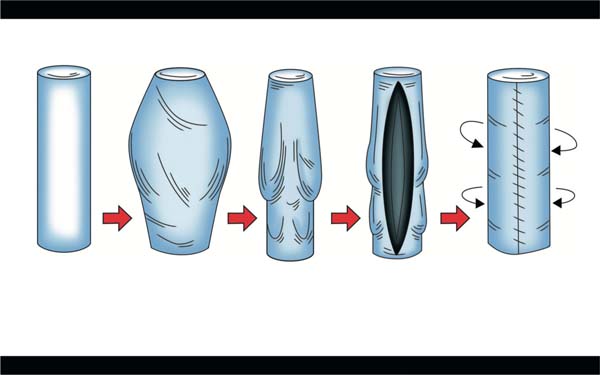
Considering the essentially cylindrical configuration of the trunk and limbs, the more pronounced appearance of “tissue descent” in certain areas results from relatively weaker local adherence zones compared with more intense ones elsewhere, reflecting different patterns of restriction to integumentary mobility. Thus, so-called horizontal or circumferential dermolipectomies promote the required tissue “suspension” (lifting effect), whereas vertical or longitudinal resections provide the equally fundamental tissue “tightening” effect (► Fig. 1). The strategic combination of suspension and tightening establishes the concept of “body readjustment,” identifying and addressing the vectors required to correct the deformities arising from this new biodynamic behavior of tissues subjected to moderate or massive weight loss.3
The literature describes several surgical strategies to achieve this body readjustment using dermolipectomies with different cutaneous traction vectors. Moreover, the resulting scars also represent an important variable in the pursuit of optimal outcomes and greater patient satisfaction.
Specifically in the trunk, the need to promote tissue tightening has guided the performance of longitudinal resections with vertical or oblique scars in association with traditional resections. In the lower body, anterior composite abdominoplasty, also known as anchor or fleur-de-lis techniques, has become increasingly popular in our practice. This surgery, either alone or combined with circumferential transverse dermolipectomy (also known as belt lipectomy), offers significant advantages, including a more refined body contour and improved waist definition. However, it does result in a more noticeable vertical scar along the midline of the abdomen.4–8
With the same goals, the lateral trunk has been approached with longitudinal resections extending from the axillary level through the flanks, achieving favorable tightening outcomes despite more apparent vertical scars.9–12 Lastly, for correction of folds and excess tissue in the posterior region, the literature classically describes dorsal “suspension” through a transverse thoracic approach with a horizontal scar positioned along the brassiere line (horizontal torsoplasty).13–17 More recently, a flank-level oblique approach, termed J-flankplasty, has been proposed to facilitate “tightening” by resecting tissue along the trunk’s longitudinal axis.18
In response to requests from multiple patients seeking correction of posterior folds and laxity, yet unwilling to accept horizontal or oblique dorsal scars, we developed a novel approach. It encompasses a posterior tissue resection via a median vertical dermolipectomy to achieve tightening and suspension through concentric advancement of the lateral flaps in a medial and cranial direction, complemented by a high horizontal dermolipectomy to adapt and redistribute the tissues recruited superiorly by cranial traction. We named this procedure vertical torsoplasty. It results in one longer longitudinal scar positioned along the posterior midline and a second, shorter transverse scar located at the transition between the cervical region and the posterior thorax. Our previous literature search in the LILACS and PubMed databases did not identify any published report to date in the medical literature describing this approach.
Objective
This study aims to present the authors’ experience in correcting dorsal laxity using vertical torsoplasty.
Methods
This open retrospective study analyzed medical records of patients who underwent vertical torsoplasty performed by the authors from March 2022 to March 2024. According to this observational design, there were no exclusion criteria, and the study included all patients—regardless of age or gender—who presented complaints of dorsal folds and laxity, and who underwent the procedure described here.
All patients received detailed information regarding the possibilities and limitations of the surgery, including the extent and positioning of the scars, potential complications, and the expected outcomes from this approach. We explained these aspects thoroughly and proceeded to the formal documentation by asking patients to sign an informed consent form. Likewise, patients provided written authorization for the use of their images for academic and scientific purposes. We submitted this study to Plataforma Brasil, under CAAE number 88882225.3.0000.5411. Moreover, the Research Ethics Committee of the Faculdade de Medicina de Botucatu, from the Universidade Estadual Paulista, approved the study.
Preoperative Marking
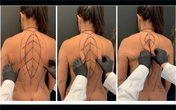
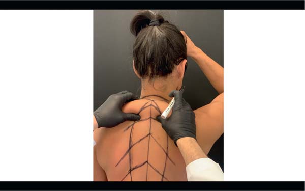
The marking design fundamentally consists of a vertical skin ellipse, whose major axis coincides with the posterior midline, more specifically, the posterior longitudinal sulcus—a linear negative contour extending from the cervical to the sacral region. With the patient in the standing position, the first marking is in the posterior midline. Next, the surgeon digitally pinches the lateral borders that will be mobilized for vertical resection performance, considering two important traction vectors toward their respective anchoring points along the midline:
Bilateral lateromedial vector, along the transverse axis, which shifts redundant dorsal skin medially from the lateral thorax toward the posterior midline, thereby promoting tissue tightening.
Caudocranial vector, along the longitudinal axis, which shifts redundant dorsal skin superiorly, opposing gravitational forces and promoting tissue suspension.
The vertical resection ellipse, incorporating both traction vectors, must be defined through simultaneous and symmetrical bidigital maneuvering, point by point, to evaluate both the tightening effect—reflected by the lateral distance between the cutaneous resection borders for approximation—and the suspension effect achieved via superior anchoring along the midline. The combined action of these vectors determines not only the amount of tissue to undergo resection but also the resulting traction direction of the operative wound edges, subsequently securing them to their respective midline anchoring points (► Fig. 2).
Depending on the degree of tissue redundancy, superior traction of the skin ellipse borders may result in tissue excess near the posterior cervical region. For this reason, the surgeon marks a compensatory dermal adipose tissue resection triangle with a high superior base, aligned with Langer’s lines between the neck and trunk. Its central vertex coincides with the midline, being positioned 2 to 3 cm short of the cervical extremity of the ellipse (► Fig. 3). This discontinuity aims to prevent convergence of the transverse and longitudinal sutures, serving as prophylaxis against wound dehiscence at a potential “T-junction” of the final scar. At the lumbar extremity of the ellipse, the marking should stop short of the sacral region, avoiding violation of this anatomical unit.

Surgical Technique
Patients underwent intravenous general anesthesia and were placed in the horizontal prone position, with all appropriate postural protective measures, in addition to thermal blanket application and pneumatic compression of the lower limbs during the anesthetic procedure. After antisepsis of the entire dorsal and lateral trunk region with 1% alcoholic chlorhexidine, the laterally redundant tissue—potentially compressed by the prone positioning—underwent traction to restore its regular mobility and reduce tension along the median suture line. The surgeon should be positioned cranial to the patient to ensure optimal control of symmetry during incision, dissection, resection, and approximation of the surgical wound edges. Additional palpation maneuvers in this position may confirm the adequacy of preoperative planning and the feasibility of approximating the marked borders (► Fig. 4A).
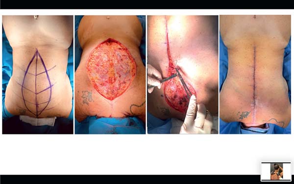
Incision of the dermal adipose tissue occurred along the entire median elliptical marking, with depth limited to a plane superficial to the muscular fascia of the superficial dorsal muscles. After bilateral incision, we proceeded to dissection and resection of the skin ellipse, preserving the underlying muscular fascia. Following removal of the vertical skin segment, the superficial anatomy of the dorsal musculature (trapezius and latissimus dorsi) became visible, and it was possible to palpate the spinous processes of the vertebral column, covered by the interspinous ligaments and central decussation of the right and left muscular fasciae (► Fig. 4B). This linear anatomical landmark serves as the anchoring point for the median suture of the lateral flaps.
After meticulous hemostasis, we proceeded to the lateral undermining of approximately 5 to 7 cm from the wound’s edges, maintaining careful preservation of the muscular fasciae to reduce tension along the suture line. This step avoids medial traction of the underlying fasciae, thereby preventing motor dysfunction and unaesthetic surface deformities or visible contour irregularities. Next, we repeated the rigorous hemostasis of the undermined areas.
Suturing of the lateral flaps began in a caudocranial direction with internal orientation stitches using 2-0 nylon, uniting the deep layer of the superficial fascia of the lateral flaps to the superior median anchoring point. This fixation involves the rigid fascio-ligamentous structure, thereby promoting the traction vectors (tightening and suspension) defined during preoperative marking (► Fig. 4C).
After establishing this initial approximation with five to six internal stitches distributed along the entire length of the ellipse—to achieve tissue tightening and suspension—we performed a supplementary continuous layered suture, incorporating the deep adipose layer and the deep median fixation line using Stratafix 0 (Medical Device Business Services, Inc.), along the entire caudo-cranial extension of the surgical wound. This deep fixation aims to preserve the posterior median longitudinal sulcus; however, its depth varies depending on subcutaneous tissue thickness, para-vertebral muscle trophism, cutaneous adherence zones, and physiological spinal curvatures.
The subdermal plane was sutured continuously with Stratafix 3-0, and the intradermal plane with Stratafix 4-0, always following the caudocranial direction (► Fig. 4D). At the conclusion of this operative stage, we assessed the amount of superior tissue redundancy and the need for transverse resection in the cervical region. In this topography, dissection must remain careful, superficial, and restricted to the subcutaneous plane, being superficial to the posterior cervical triangle, where the skin is thinner and adherent to the trapezius fascia, which requires preservation. This precaution prevents troublesome bleeding and reduces the risk of segmental injury to the accessory nerve, whose motor component innervates the sternocleidomastoid and trapezius muscles, thereby contributing to head rotation and stability, as well as elevation of the scapulae, arms, and shoulders.
As described in the preoperative marking, the orientation of the base and final scar of the cervical triangle for resection should coincide with Langer’s lines at the posterior cervicothoracic transition. After dermal adipose tissue resection at this level, we performed careful hemostasis, avoiding thermal injury to the underlying accessory nerve, followed by continuous layered closure using the same sutures previously described. All sutures received a complementary adhesive application (Dermabond Prineo glue, J&J MedTech) and a secondary occlusive dressing.
Postoperative Follow-up and Evaluation
Patients underwent periodic follow-up visits for dressing changes, serial photographic documentation, and guidance regarding rest and resumption of routine activities. Retrospective analysis of all medical records documented the occurrence, progression, and consequences of complications.
Outcome quality was assessed retrospectively using standardized photographs taken 3 to 12 months postoperatively. This subjective evaluation was performed by the authors and by an independent observer, a member of the nursing staff. Scoring aimed to demonstrate professional satisfaction with the improvement in body contour achieved at the conclusion of treatment, using a simple visual analog scale (VAS) categorized as unsatisfactory, fair, good, or excellent.
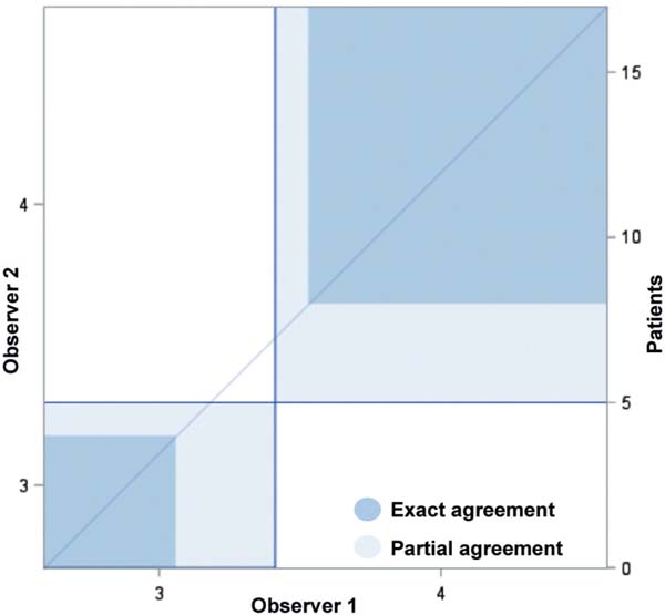
Interobserver agreement between the two healthcare professionals was evaluated using Cohen’s kappa coefficient, with the SAS software (Sistema de Análise Estatística, SAS Institute), version 9.4.
Results
A total of 17 patients underwent surgery during the study period, and their medical records were reviewed retrospectively. The cohort comprised 16 women and 1 man, with a mean age of 37 (range: 29–54) years. All patients presented with preoperative complaints of dorsal laxity characterized by excess tissue and folds. There were 4 cases (23.5%) classified as Pittsburgh grade 1 (adiposity), and 13 (76.5%) as grade 2 (cutaneous folds).19
A prior history of negative weight variation ranging from 9 to 50 (mean: 20) kg was observed, with 3 patients (17.6%) having undergone bariatric surgery and 14 (82.3%) losing weight through diet and exercise. The mean preoperative body mass index (BMI) was 27 (range: 26–29). Most patients had no previous scars or surgical procedures in the dorsal region, except for one subject who had previously undergone classical horizontal torsoplasty and still reported discomfort and residual laxity at the site. Patients’ general conditions were analyzed through clinical and laboratory evaluations, which revealed no relevant changes or contraindications to surgery.
The mean operative time for dorsal vertical torsoplasty was 90 (range: 70–120) minutes. Some patients underwent additional procedures, which were not included in the operative time analysis. No relevant intraoperative anesthetic or surgical complications were observed. Postoperative recovery was uneventful, and all patients were discharged after a mean hospital stay of 30 hours (range: 24–48 hours).
Postoperative follow-up ranged from 3 to 12 months, during which patients underwent serial clinical evaluations and standardized photographic documentation. ► Table 1 summarizes postoperative complications. No major adverse events, including infection, necrosis, hematoma, or thromboembolic events, were observed.
| Number | Gender | Complication | Assessment 1 | Assessment 2 |
|---|---|---|---|---|
| 1 | Female | None | Excellent | Excellent |
| 2 | Female | None | Excellent | Good |
| 3 | Female | None | Good | Excellent |
| 4 | Female | None | Good | Good |
| 5 | Female | Seroma and dehiscence | Good | Good |
| 6 | Female | Dehiscence and enlarged scar | Good | Good |
| 7 | Female | None | Excellent | Excellent |
| 8 | Female | Enlarged scar | Good | Good |
| 9 | Female | None | Excellent | Excellent |
| 10 | Female | None | Good | Excellent |
| 11 | Female | None | Excellent | Excellent |
| 12 | Female | None | Excellent | Excellent |
| 13 | Female | None | Excellent | Excellent |
| 14 | Female | None | Excellent | Excellent |
| 15 | Female | None | Excellent | Excellent |
| 16 | Male | None | Excellent | Excellent |
| 17 | Female | Enlarged scar | Good | Excellent |

We observed two cases of wound dehiscence (7–8cm), which occurred on postoperative day 7. Initial management consisted of serial dressings followed by resuturing on postoperative day 15, with satisfactory early healing. However, both cases subsequently developed scar widening, requiring surgical scar revision 6 months after resuturing. Furthermore, there was one case of seroma occurred in the interscapular region approximately 20 days postoperatively. It progressed to spontaneous drainage and formation of a 2cm orifice secondary to localized dehiscence along the median scar. Surgical management included cavity exploration, hemostasis, wound edge reapproximation, resuturing with fixation of the lateral flaps to the deep planes, and application of negative-pressure wound therapy over the revised 15-cm scar.
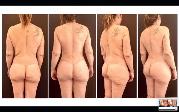
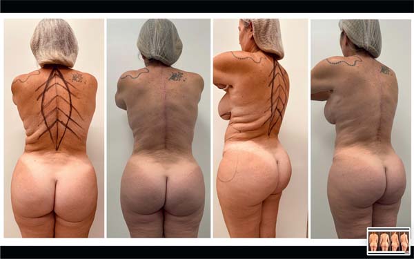
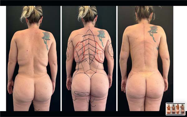
Most cases incorporated additional body contouring procedures into the surgical plan, with variable impact on the isolated evaluation of outcomes attributable solely to vertical torsoplasty. Analysis of serial photographs documenting clinical progression and assessment of outcomes achieved with the employed technique yielded high and concordant subjective satisfaction scores from both the author and the independent observer (► Figs. 5–7). Although the sample size was relatively small (n = 17), the kappa coefficient of 0.4925 indicated moderate interobserver agreement (► Graph 1).

Discussion
Surgical management of postweight loss skin laxity is traditionally performed through dermolipectomies, in which preoperative planning determines both the extent of tissue resection and the vectors of flap mobilization. In this context, trunk contour restoration requires a combination of suspension and tissue-tightening maneuvers, resulting in horizontally and/or vertically oriented scars along the trunk.3
Vertical scars, whether anterior or lateral, although more evident and often associated with a negative aesthetic perception, result in improved and desirable adaptation of the skin envelope to the cylindrical thoracoabdominal framework. In the dorsal region, however, unlike the anterior and lateral midlines, there is a pronounced zone of cutaneous adherence to deeper structures, forming a distinct negative contour characterized by the posterior median sulcus. This anatomical element is considered fundamental by some authors, who even advocate sparing these structures during horizontal or oblique dorsal dermolipectomies to maintain the nuances of beauty and natural appearance in this region.20 Perhaps due to this perception, no description of vertical approaches for treating posterolateral laxity has been identified in medical literature.
From our perspective, positioning a vertical scar along the posterior median sulcus enabled more appropriate vectors for tissue readjustment and resulted in a scar better concealed within a natural fold or groove of the body. The primary challenge of this approach, however, seemed to be preservation of surface anatomy, avoiding effacement of the posterior median sulcus and potential loss of osteomuscular definition in the three-dimensional plane. Moreover, correction required lateromedial tightening and effective cranial suspension of the tissues. Accordingly, the direction of approximation of the lateral borders of the posterior vertical ellipse was specifically planned to generate both tightening and suspension vectors, with anchorage in deeper planes coinciding with the posterior midline.
In a substantial portion of our cases, bilateral superior traction recruited tissue toward the posterior cervical base. For this reason, we advocated decomposition of this high thoracic redundancy by resecting an “inverted triangle” with a cervical base, whose removal and closure position a well-camouflaged horizontal scar between the neck and upper thorax.
In our series, resection of this superior compensatory triangle was indicated in most cases, depending on the magnitude of dorsal redundancy and tissue accumulation at the superior apex of the vertical ellipse. Whenever possible, maintaining a cutaneous bridge between the vertical scar and the superior horizontal scar helps prevent wound dehiscence related to a potential “T-junction” of the three posterior flaps.
To some extent, and in an inverted manner, the vertical torsoplasty strategy reproduces the vectors from anchor abdominoplasty, promoting readjustment through a combination of suspension and posterolateral tightening. In our experience, the posterior vertical approach was performed in early and intermediate cases of dorsal laxity (Pittsburgh grades 1 and 2). Furthermore, marking maneuvers and execution of the surgical strategy were relatively simple and reproducible, with a low rate of minor complications and complete absence of major adverse events. In nearly all cases, it was possible to correct lateral-dorsal laxity and folds extending from the flanks to the posterolateral region, achieving good positioning and quality of the median vertical scar while preserving the aesthetic and anatomical integrity of the back, with well-defined structural contour at the level of the posterior median sulcus.
This study aimed to retrospectively present findings from the authors’ initial experience with a specific posterior vertical dermolipectomy approach not previously described, at least within the literature consulted. As a limitation of our method, we recognize that the number of patients remains limited, precluding more definitive conclusions, including regarding the potential effectiveness of this approach in more severe cases of dorsal laxity (Pittsburgh grade 3), since such patients were not included in our series. Likewise, further studies are needed to better define indications, complications, and the true applicability of this technique, including longer postoperative follow-up, incorporation of scientifically validated patient satisfaction measures, and controlled comparisons with outcomes achieved using alternative approaches previously described in the literature.
Conclusion
The vertical torsoplasty strategy presented for the treatment of patients with mild to moderate dorsal laxity demonstrated itself to be a simple, safe, and reproducible procedure, with favorable scar positioning and quality. It achieves trunk contour restoration through combined tissue tightening and suspension while preserving the anatomical definition of the structures composing the posterior median sulcus.
REFERENCES
1. Mendes F, Viterbo F, Gaiotto JA. Particularizando as deformidades no paciente pós-bariátrico. In: Mendes FH, Viterbo F, editores. Cirurgia plástica pós-bariátrica. Rio de Janeiro: Di Livros; 2016:110–128
2. Strauch B, Herman CK. Describing the Deformities. In: Rubin JP, Jewell ML, Richter DF, Uebel CO, editores. Body Contouring and Liposuction. Filadélfia: Elsevier/Saunders; 2012:580–584
3. Mendes F, Viterbo F, Alves AL. Reajuste corporal pós-bariátrico – conceitos e tendências. In: Mendes FH, Viterbo F, editores. Cirurgia plástica pós-bariátrica. Rio de Janeiro: Di Livros; 2016:87–101
4. Costa LF, Manta AM, França AS, Cavalcante HA, Nahon M. Abdominoplastia vertical modificada em pacientes ex-obesos. Rev Soc Bras Cirurgia Plast 2003;18(03):71–74. Disponível em: https://www.rbcp.org.br/Content/imagebank/pdf/18-03-07-pt.pdf
5. Kaluf K, Salgado AMF. Abordagem anterior composta. In: Mendes FH, Viterbo F, editores. Cirurgia plástica pós-bariátrica. Rio de Janeiro: Di Livros; 2016:292–300
6. Cintra W Jr, Modolin M, Rocha RI, Gemperli R. Abordagem circunferencial. In: Mendes FH, Viterbo F, editores. Cirurgia plástica pósbariátrica. Rio de Janeiro: Di Livros; 2016:301–318
7. Mendes FH, Viterbo F, Luna ALAP. Inner scar umbilicus: New horizons for vertical abdominoplasty. Plast Reconstr Surg 2018; 141(04):507e–516e. Doi: 10.1097/PRS.0000000000004258
8. Mendes FH, Viterbo F, Moragas WR. Finesse in fleur-de-lis abdominoplasty. Clin Plast Surg 2024;51(01):81–93. Doi: 10.1016/j.cps.2023.09.001
9. Pianguy I. Correction of lipodystrophy of the lateral thoracic aspect and inner side of the arm and elbow dermosenescence. Clin Plast Surg 1975;2(03):477–483
10. Clavijo-Alvarez JA, Hurwitz DJJ. J torsoplasty: a novel approach to avoid circumferential scars of the upper body lift. Plast Reconstr Surg 2012;130(02):382e–383e. Doi: 10.1097/PRS.0b013e31825903e5
11. Richter DF, Stoff A. Upper Body Lift with Lateral Excision. In: Rubin JP, Jewel ML, Richter DF, Uebel CO, editores. Body Contouring and Liposuction. Filadélfia: Saunders/Elsevier; 2012:167–175
12. Rahban SR, Gross JE. A new approach to correction of truncal redundancy after massive weight loss–the lateral thoracoabdominoplasty. Aesthet Surg J 2007;27(05):518–523. Doi: 10.1016/j.asj.2007.06.003
13. Richter DF, Stoff A. Back and Lateral Fold Contouring. In: Nahai F, editor. The Art of Aesthetic Plastic Surgery: Principles and Techniques. 2nd ed. St. Louis: Quality Medical Publishing; 2010
14. Hunstad JP, Repta R. Bra-line back lift. Plast Reconstr Surg 2008; 122(04):1225–1228. Doi: 10.1097/PRS.0b013e3181858fa4
15. Rubin JP, Michaels J. Upper Body Lift for Correction of Back Rolls. In: Strauch B, Herman CK, editores. Encyclopedia of Body Sculpting After Massive Weight Loss. Nova York: Thieme; 2011:224–229
16. Strauch B, Rohde C. Back Contouring. In: Strauch B, Herman CK, editores. Encyclopedia of Body Sculpting After Massive Weight Loss. Nova York: Thieme; 2011
17. Hunstad JP, Knotts CD. Transverse upper body lift. In: Rubin JP, Jewel ML, Richter DF, Uebel CO, editores. Body Contouring and Liposuction. Filadélfia: Saunders/Elsevier; 2012:159–165
18. Hurwitz DJ, Beidas O, Wright L. Reshaping the oversized waist through oblique flankplasty with lipoabdominoplasty. Plast Reconstr Surg 2019;143(05):960e–972e. Doi: 10.1097/PRS.0000000000005574
19. Song AY, Jean RD, Hurwitz DJ, Fernstrom MH, Scott JA, Rubin JP. A classification of contour deformities after bariatric weight loss: the Pittsburgh Rating Scale. Plast Reconstr Surg 2005;116(05): 1535–1554; discussion1545–1546. Doi: 10.1097/01.prs.0000182606.92069.13
20. Hurwitz DJ. Aesthetic refinements in body contouring in the massive weight loss patient: trunk. Plast Reconstr Surg 2014; 134(06):1185–1195. Doi: 10.1097/PRS.0000000000000759
1. Plastic Surgery Department, Private Practice, São Paulo, SP, Brazil
2. Plastic Surgery Program, Faculdade de Medicina de Botucatu, Universidade Estadual
Paulista (UNESP), Botucatu, SP, Brazil
Financial Support
Address for correspondence Flavio Henrique Mendes, Disciplina de Cirurgia Plástica, Faculdade de Medicina de Botucatu, Universidade Universidade Estadual Paulista (UNESP), Botucatu, SP, Brazil Estadual Paulista (UNESP), Avenida Professor Mário Rubens Guimarães Montenegro s/n, Botucatu, SP, CEP: 18618–687, Brazil (e-mails: mendesmd@fhmendes.com.br; flaviomendesmd@gmail.com).
Article received: December 19, 2024.
Article accepted: December 14, 2025.
Conflict of Interests The authors have no conflict of interests to declare.
 Read in Portuguese
 Read in English
 PDF PT
 Print
Print
 Send this article by email
 How to Cite
 Mendeley
 Pocket
 Facebook
 Twitter